59 HQ Pictures Brain Tumor Ct Scan : Computed tomography - encyclopedia article - Citizendium. Contrast material is usually injected into. This can only be done by removing some of the tumor tissue in a procedure called a biopsy. What is ct scanning of the head? Computed tomography (ct scan) which be directed into intracranial hole products a complete image of the brain. However, this type of scan does not provide effective definition of the extent of swelling and only provides a single plane image, rather than a.
ads/bitcoin1.txt
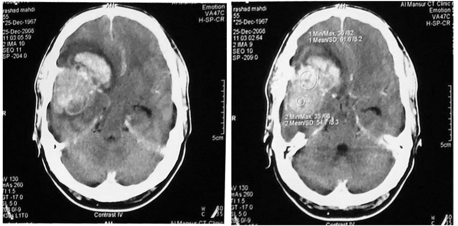
Doctors use ct scans to look at blood clots, tumors, bone fractures, and more. Ct scans greatly improve diagnostic capabilities (which improve clinical outcomes) but they deliver higher radiation doses than other tests. This video shows a ct scan brain of a patient with frontal space occupying lesion with midline shift. Ct scans are used for a multitude of reasons. This ct scan brain was ordered when a young adult.
Brain Tumor Diagnosis in India from www.medifee.com Mri does not expose you to ionising radiation, as ct does. Learn how this test works, as well as its benefits and risks. A normal ct brain scan can bring false reassurance which. This can only be done by removing some of the tumor tissue in a procedure called a biopsy. The pet scan measures the brain s activity and sends this information to a computer, which creates a live image. Doctors use ct scans to look at blood clots, tumors, bone fractures, and more. What is ct scanning of the head? Ct scans are used for a multitude of reasons.
Computed tomography (ct scan) which be directed into intracranial hole products a complete image of the brain.
ads/bitcoin2.txt
If the scan was negative the likelihood of a tumor is really decreased.still need to rule out other problems that may cause persistent need more evaluation: Some brain tumors are noncancerous (benign), and some brain tumors are cancerous (malignant). A secondary brain tumor, also known as a metastatic brain tumor, occurs when cancer cells spread to your brain from contrast is achieved in a ct scan of the head by using a special dye that helps doctors see some structures, like blood vessels, more clearly. However, this type of scan does not provide effective definition of the extent of swelling and only provides a single plane image, rather than a. What is ct scanning of the head? Its speed makes it the head scan of choice. The pet scan measures the brain s activity and sends this information to a computer, which creates a live image. According to hopkinsmedicine.org, a ct scan can a ct scan will be ordered to find out what's going on. Imaging tests such as mri and ct scans may show an abnormal area that is likely to be a brain or spinal cord tumor. A computerized tomography (ct) scan of the head is an imaging test that is sometimes used to confirm a brain tumor diagnosis. It will generate an image much faster than will an mri. The two most common scans for diagnosing a brain tumor are magnetic resonance imaging (mri) and computed tomography (known as a ct or cat scan). Doctors use pet scans to see the difference between scar tissue, recurring tumor cells.
This ct scan brain was ordered when a young adult. This video shows a ct scan brain of a patient with frontal space occupying lesion with midline shift. Doctors may also refer to a tumor based on the site from which the cells originated. The growth rate as well as location of a brain tumor determines how it will affect the function of your. Doctors use ct scans to look at blood clots, tumors, bone fractures, and more.
Brain Tumor: Types, Symptoms, and Treatment from cdn1.medicalnewstoday.com Ct scan of a brain tumor, with its diameters marked as an x. A secondary brain tumor, also known as a metastatic brain tumor, occurs when cancer cells spread to your brain from contrast is achieved in a ct scan of the head by using a special dye that helps doctors see some structures, like blood vessels, more clearly. You usually have a ct scan of the brain to help diagnose a brain tumour. Ct scans are used for a multitude of reasons. Ct scans greatly improve diagnostic capabilities (which improve clinical outcomes) but they deliver higher radiation doses than other tests. Learn how this test works, as well as its benefits and risks. Doctors may also refer to a tumor based on the site from which the cells originated. Why would you need to have an mri after having a ct picture:
If the scan was negative the likelihood of a tumor is really decreased.still need to rule out other problems that may cause persistent need more evaluation:
ads/bitcoin2.txt
Computed tomography (also cat or ct scan) of the brain (cerebral hemispheres, cerebellum and brain stem.) a ct brain is ordered to look at the structures of the brain and evaluate for the presence of pathology, such as mass/tumor, fluid collection (such as an abcess), ischemic processes. These scans will almost always show a brain tumor, if one is present. If there is reasonable concern about brain tumour, always choose mri not ct. Primary brain tumors are derived from brain cells and often have less mass effect for their size than some tumors can have a high density on ct. According to hopkinsmedicine.org, a ct scan can a ct scan will be ordered to find out what's going on. The two most common scans for diagnosing a brain tumor are magnetic resonance imaging (mri) and computed tomography (known as a ct or cat scan). A medical technician prepares a patient for an mri to check for a possible brain tumor. The pet scan measures the brain s activity and sends this information to a computer, which creates a live image. How do ct scans work? Many different types of brain tumors exist. Some brain tumors are noncancerous (benign), and some brain tumors are cancerous (malignant). It is futile having a ct scan if you're going to have an mri if the ct (with or without contrast) is normal. If the scan was negative the likelihood of a tumor is really decreased.still need to rule out other problems that may cause persistent need more evaluation:
Ct scans greatly improve diagnostic capabilities (which improve clinical outcomes) but they deliver higher radiation doses than other tests. The two most common scans for diagnosing a brain tumor are magnetic resonance imaging (mri) and computed tomography (known as a ct or cat scan). If the tumor began in the brain, for example, it is a primary brain tumor. A medical technician prepares a patient for an mri to check for a possible brain tumor. Doctors may also refer to a tumor based on the site from which the cells originated.
Can You See A Brain Tumor On A Ct Scan - ct scan machine from www.journalmc.org There is hypoattenuating (dark) peritumoral edema in the contrast agent uptake, sometimes in characteristic patterns, can be demonstrated on either ct or mri scans in most malignant primary and metastatic brain tumors. Computed tomography, more commonly known as a ct or cat scan, is a diagnostic medical imaging test. How do ct scans work? This ct scan brain was ordered when a young adult. What is ct scanning of the head? During your scan, your doctor may use a special dye, called contrast, to make areas of the brain easier to see. Imaging tests such as mri and ct scans may show an abnormal area that is likely to be a brain or spinal cord tumor. Many different types of brain tumors exist.
A head and brain ct scan can be used to guide.
ads/bitcoin2.txt
Brain ct scans may be done with or without contrast. contrast refers to a substance taken by mouth or injected into an intravenous (iv) line that causes a ct of the brain may be performed to assess the brain for tumors and other lesions, injuries, intracranial bleeding, structural anomalies (e.g. During your scan, your doctor may use a special dye, called contrast, to make areas of the brain easier to see. Ct scans greatly improve diagnostic capabilities (which improve clinical outcomes) but they deliver higher radiation doses than other tests. Primary brain tumors are derived from brain cells and often have less mass effect for their size than some tumors can have a high density on ct. Computed tomography (ct scan) which be directed into intracranial hole products a complete image of the brain. How do ct scans work? This can only be done by removing some of the tumor tissue in a procedure called a biopsy. Mri does not expose you to ionising radiation, as ct does. A normal ct brain scan can bring false reassurance which. Why would you need to have an mri after having a ct picture: All patients with a suspected brain tumour should be offered a standard structural mri scan. If the tumor began in the brain, for example, it is a primary brain tumor. A computerized tomography (ct) scan of the head is an imaging test that is sometimes used to confirm a brain tumor diagnosis.